
Model Card
An Image Classifier that predicts the presence of certain Brain tumours from their MRI scans
Model Details
A 134M Parameter ConvNet designed for classification of Brain tumours in MRI scans.
Paper
Interpretable Deep Learning for Brain Tumor Diagnosis: Occlusion Sensitivity-Driven Explainability in MRI Classification DOI: 10.21015/vtse.v13i2.2082
Uses
Direct Use
Load the model, finetune the model if needed or just go straight towards generating inferences using the model.
Downstream Use
Finetune the model on other diagnostic scans, though the model only accepts grayscale images of size 256x256.
How to Get Started with the Model
![]()
Training
The colab notebook used to train the model can be found below
![]()
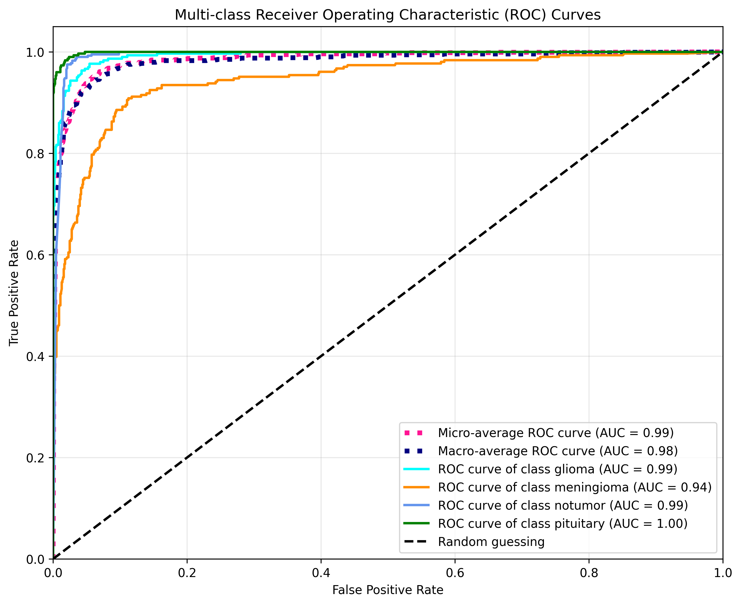
Evaluation
Metrics
| Class | Precision | Recall | F1-Score | Support |
|---|---|---|---|---|
| Glioma | 0.96 | 0.87 | 0.91 | 300 |
| Meningioma | 0.84 | 0.71 | 0.77 | 306 |
| No Tumor | 0.88 | 1.00 | 0.93 | 405 |
| Pituitary | 0.93 | 0.99 | 0.96 | 300 |
| Accuracy | 0.90 | 1311 | ||
| Macro Avg | 0.90 | 0.89 | 0.89 | 1311 |
| Weighted Avg | 0.90 | 0.90 | 0.90 | 1311 |
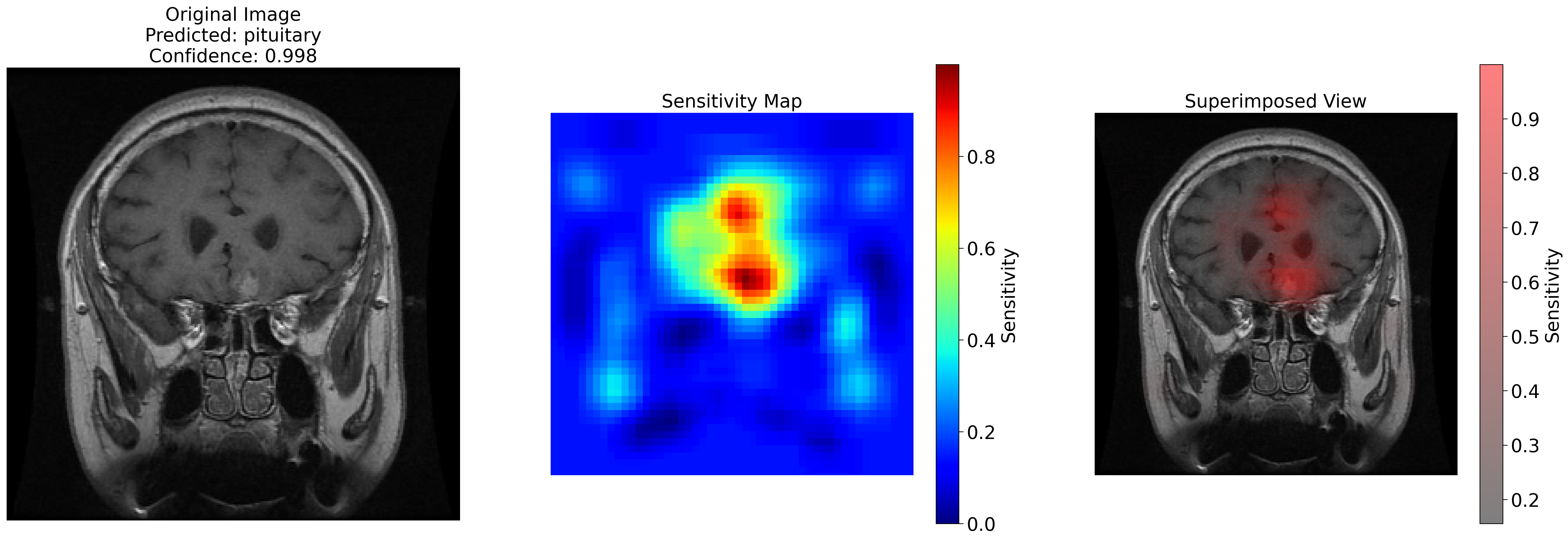
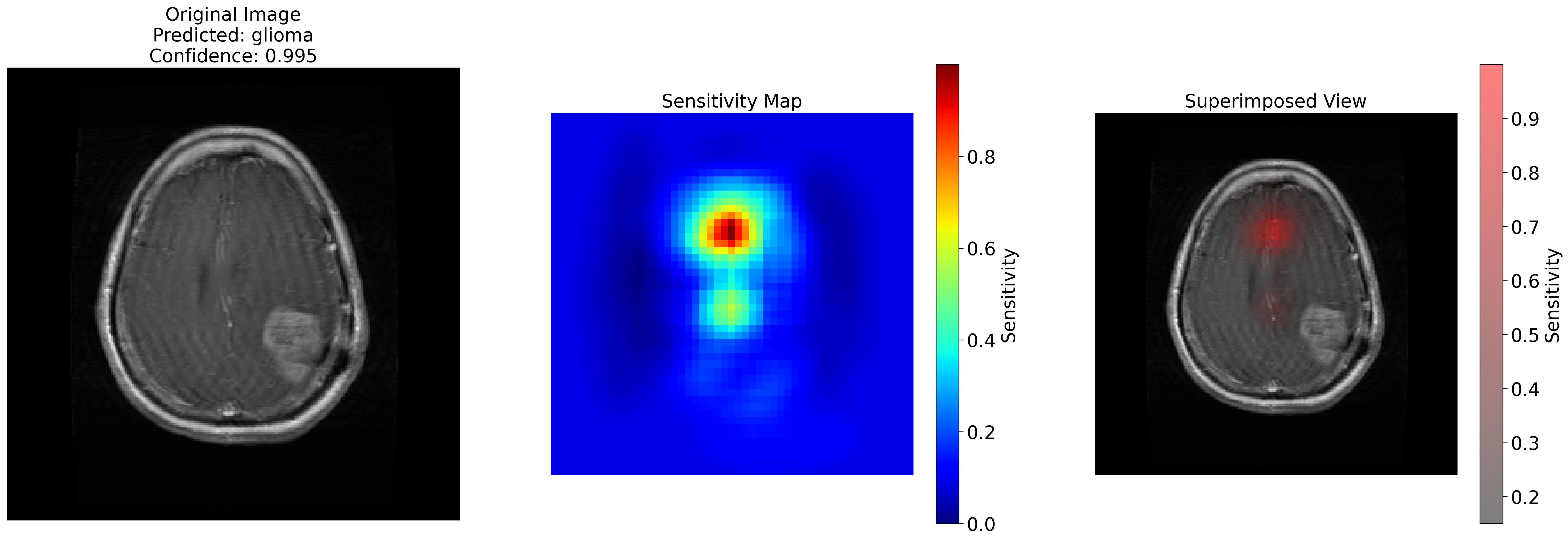
Results
This model was developed for my project that can be found on github here
. This project involved generating sensitivity maps to explain the predictions of the model.
These maps assign values to areas of the image that act as feature importance markers.
- Downloads last month
- 27